

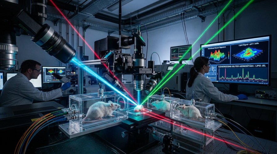
Scientists have combined laser-based imaging with gene activity mapping to spot senescent cells, the ageing cells linked to tissue decline, in mouse lung and skin. They found a fat-linked “vibrational” signal that appears to mark them across both tissues.
Lead: Ageing cells are hard to catch. A new technical report in Nature Aging, published on 21 September 2026, describes a way to find them by shining light on tissue and reading how its molecules vibrate, no destruction required. In mouse lung and skin, the team found a shared chemical signal that flagged senescent cells.
The problem with spotting “old” cells
Senescent cells, often nicknamed “zombie cells”, are cells that have stopped dividing but linger in tissue. They are tied to ageing and to how tissues repair themselves. The trouble, according to the authors, is that they are usually defined by single molecular markers, which hides how a cell’s chemistry, gene activity and structure change together.
Enter RamanOmics
The team’s answer is RamanOmics. It combines label-free hyperspectral Raman imaging, which reads the chemical makeup of tissue from the way its molecules interact with light, with two gene-activity techniques: single-nucleus RNA sequencing and spatial transcriptomics. Together they link a cell’s biochemical state to its gene programs, at single-cell resolution and with its position in the tissue preserved.
Lung and skin age differently
The researchers applied the method to young and old mouse tissue. In the lung, senescent cells were enriched for extracellular matrix remodelling, the reworking of the scaffolding around cells, and for transforming growth factor-β signalling. In the skin, senescence was dominated by genes involved in epidermal differentiation, including Krt10, Lor and Sbsn.
One shared clue
Despite those differences, one feature held across both tissues: a lipid-linked Raman signature, in the range of 1,131 to 1,135 cm⁻¹, that marked cells positive for p21, a well-known senescence marker. Using machine learning, the team built a multimodal “barcode” that can identify senescent cells in place, without destroying the sample.
A twist in wound healing
The method also revealed something in a mouse wound-healing model. Senescent cells there reactivated the same epidermal differentiation genes (Krt10, Lor and Sbsn), alongside stronger lipid-associated Raman signatures. That points to senescent cells playing an active role in repair, not just in ageing.
What comes next
The authors describe RamanOmics as a tissue-agnostic framework for scalable, multimodal profiling of cellular states. Note the limits: this work was done in mice, in lung and skin, and the report does not claim results in people. But a non-destructive way to see ageing cells in their natural setting could give longevity researchers a sharper tool for asking where these cells sit, what they are doing and how they change during repair.
Source:
https://www.nature.com/articles/s43587-026-01219-7
Transparency Note: This article was prepared with the assistance of artificial intelligence technologies. Every piece is thoroughly reviewed and verified by our editorial team, who retain full responsibility for the final content.
Picture: Gemini AI